
Thursday 28 August 2025
On Monday 18 August, a small group of medically interested physics students ventured over to experience the scientific wonder of modern radiology equipment. As we entered the grand doors of Middlemore Hospital, we were greeted by some lovely staff who were to take us around on an exclusive tour of the hospital and its various equipment. After a short introduction, we were off like electrons accelerating through a cathode ray tube. 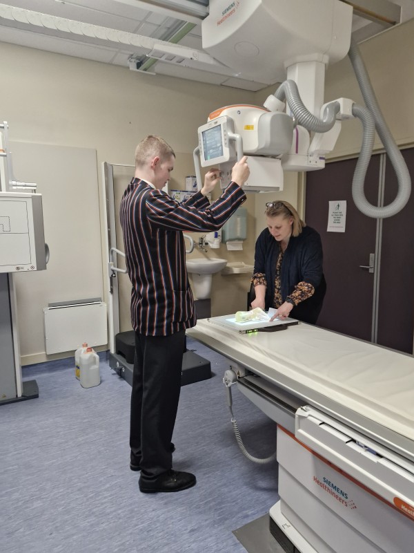
As we travelled through the bustling corridors of the hospital, our first stop was the magnificent MRI machines. Mega-magnets of 1.5 and 3 Teslas were on full display, and a demonstration of their magnetic power involving metal balls shooting across the room was truly awesome. Having the opportunity to ask questions to an experienced radiologist was a privilege, and our brains absorbed all the new fascinating information. Bursting through the doors like X-rays through tissue with a low absorption coefficient, our next stop was the X-ray Department.
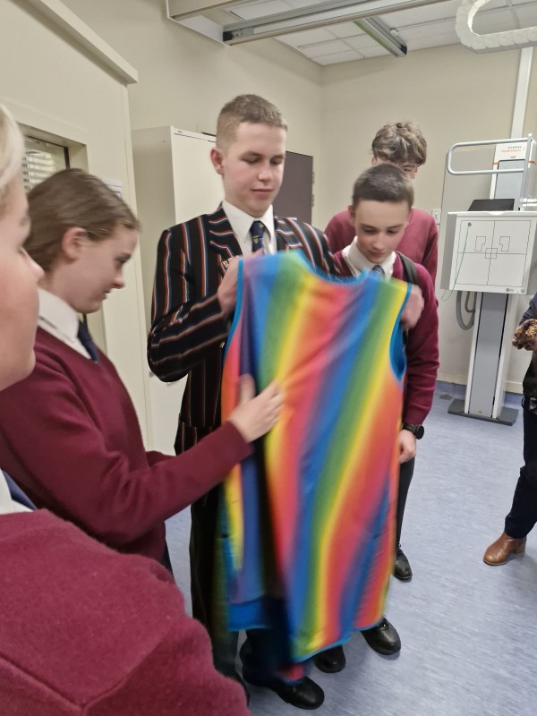
Walking into a lead-lined room, we were ‘lead’ along to the X-ray machine. After some general information of the inner machinations of the device, as well as the various methods of detection, due to a staff shortage, the King’s College Middlemore trip team had to perform a vital X-ray on a decapitated hand. After a particularly handsome student primed the machine, we went back behind the control panel and saw an image of the hand! It was x-ray ordinary. We learned about the different effects of voltage power and imaging technology. Afterwards, we went to the Ultrasound Department, not because we were all pregnant (although I did have a rather large lunch beforehand! After another demonstration, we were told about the advantages of ultrasound, the use of the gel and the various different settings that could be employed, which was truly eye opening.
 Our final stop was the CT scanner. Once again, we were lucky enough to talk to two radiologists about the uses and workings of the CT scanner, and how it created slices of scans of our body to piece together a comprehensive 3D picture. C-T-riffic! We learnt about iodine tracer and then got to see the various gizmos and gadgets associated with the machine. Saying our goodbyes to the staff, we headed out of the hospital, having our final dis-charge from the Radiology Department, excited like photoelectrons.
A sincere thank you to all the staff from Middlemore Hospital for this fascinating experience.
Charlie Wadham (Year 13, Averill)